Articles

Is using ear prosthetic to cover handicapped ear as an appropriate solution?
A silicon prosthetic ear can be created to look very similar to a patient’s normal ear. The ear prosthesis procedure is simpler and less risk than . It provides an option for patients who are not candidates for surgery. A disadvantage of prosthetic ear is the fading of ear color and deterioration of the prosthesis requires replacement every 2–3 years. There are two methods that are used to attach the ear prostheses. One uses a glue to adhere the ear to the skin. The second method uses titanium implants which are placed onto the bone around the ear. The prosthetic ear can then be “snapped” into place. The prosthetic ear using a glue needs to be removed at night and replaced each morning, difficulty hiding the seam between the prosthesis and cheek normal skin and may lose the prosthesis during sport or rough play. The prosthetic ear using implants do not to be removed and replaced each day.

Can Microtia be detected in an ultrasound?
No. Typically, sonographers do not focus on looking for Microtia or the details of the ears. Sonographers focus more on the organs of the baby, to make sure everything is developing correctly. Ultrasounds are used to detect abnormal features such as in Down’s Syndrome or mis-shapen limbs, along with general blood screening if requested, etc…. It is possible that you can ask the sonographer to look for more details as ultrasound technology today has improved greatly. However, this would only notify you that your child would have Microtia. An ultrasound can not prevent Microtia from happening nor can it fix it.

What is surgery to shape the microtia (defective ear) using a plastic frame?
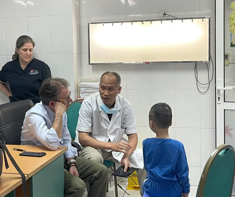
Microtia plastic surgery using a is also called . First introduced by Dr. John Reinisch, Medpor is a surgery that uses a foam ear frame with autologous tissue to shape the ear. The polyethylene frame is sculpted to resemble a healthy ear frame (Figure 1) is implanted in the appropriate position (Figure 2) and then covered by a thin layer of tissue that turns down from the subcutaneous layer (called the temporal flap) and cover with a skin graft taken from patient’s own thigh or abdomen. All of these steps are performed in one operation. Advantages: Can be done with three-year-olds, before they go to school; For children with hearing loss, surgery to restore the ear canal can be done before or at the same time with reconstructing the defective ear; Minimizes pain relief while allowing children to go home (outpatient) one hour after surgery; Reconstructive surgery is performed once and in 80% of cases are outpatient; The reconstructed ear is formatted to correspond to the normal ear in size and appearance (Figure 3); The reconstructed ear can withstand most sports. Disadvantages: The ear frame is the plastic that replaces the body’s tissue; Only a few surgeons are trained in this surgery, an inexperienced ear surgeon with a defective ear will lead to unexpected results. In Vietnam, Dr. Nguyen Hong Ha, after a period of practice under the direct guidance of Dr. John Reinisch and his associates of Faculty of Facial Surgery, Plastic and Aesthetics, successfully performed the first Medpor ear reconstruction in Vietnam at Viet Duc Friendship Hospital. Photographs 2 months after surgery show that the patient has had wonderful reconstructive ears.

Why is ear-shaping surgery with endoscopic-assissted superiorto open surgery?
(one-time surgery) using (Medpor method) or using as performed by Associate Professor Dr. Nguyen Hong Ha and colleagues at Vietnam-GermanyFriendship Hospital are requires the use of to cover the cartilage frame. In order to harvest the shallow temporoparietal fascia tissue flap, attention should be paid to the superficial temporoparietal vascular system and temporoparietal nerve. The temporoparietal vascular system comes out from the upper surface of the parotid gland and goes along with the temporoparietal nerve (see figure 1). The superficial temporal artery originates from the external carotid artery. This artery is often the cause of bleeding during surgery. The superficial veins are outside and often behind the artery. The temporoparietal nerve is parallel to it behind the artery. The temporoparietal nerve receives sensations from the ear canal, the outer ear canal, the eardrum, and the skin of the temporoparietal region. Just in front of the ear, it divides the extremities into the skin of the temporoparietal region (see figure 1). In surgery without endoscope-supported, surgeons make a long incision to open the dissection cavity and observe the flap details with the naked eye. This technique is less precise than endoscopic technique and the risk are cutting to the artery and vein and also the long keloid scare alopecia (Fig. 2) In surgery with supportive endoscopy, a small incision is made to insert the camera endoscope into the dissection cavity. The image inside the dissection cavity sent by the receiver at the end of the scope is enlarged on a large screen for good viewing, so the extraction is performed accurately. Thanks to that, the flap of the temporoparietal connective tissue with the vessels ensures good blood supply and circulation, less damage to the temporoparietal nerve while minimizing incision, scar size and hair loss (Fig 3). # One-stage ear-shaping surgery# Associate Professor Nguyen Hong Ha# Vietnam-Germany Friendship Hospital# open ear-surgery# endoscopic-assisted ear-surg

Why Do Some Patients with Congenital Microtia Choose Ear Reconstruction Surgery After Using a Silicone Prosthetic Ear?
For patients with congenital microtia , wearing a silicone prosthetic ear offers a major advantage: they can achieve a natural-looking ear without undergoing complex surgery. However, this solution also comes with several challenges: 👉 Aesthetic limitations – The prosthesis may not perfectly match the skin tone, and the implant base can sometimes be visible. While a skilled prosthetist can improve these issues, they are not always completely resolved. 👉 Social discomfort – In certain situations, patients need to remove their prosthetic ear, such as during sports, swimming, gym workouts, or other physical activities. This can cause embarrassment, self-consciousness, and social anxiety . 👉 Lack of naturalness and permanence – Even with modern techniques, a prosthetic ear cannot fully provide the sense of confidence and authenticity that comes with a surgically reconstructed ear made from the patient’s own tissue. Because of these reasons, many patients eventually seek ear reconstruction surgery for a more natural, stable, and lifelong solution. Today, thanks to advances in medical techniques, particularly the one-stage, single-incision endoscopic ear reconstruction performed by Associate Professor Nguyen Hong Ha , patients can achieve an ear that looks natural, symmetrical, and long-lasting—helping them regain confidence in daily life and social interactions. Please send us a message via our website or call our hotline at +84 974.700.600 to book an appointment.

Atresia Repair Surgery Can Restore Hearing and Transform Lives
For children born with Aural Atresia —a condition where the ear canal is absent or underdeveloped—hearing loss can make everyday listening a challenge. This is especially true in noisy environments such as classrooms, restaurants, playgrounds, and cars, where background noise makes it hard to identify where sounds are coming from. “Sound localization is essential for safety, communication, and learning,” explains ear specialists. “When one ear is affected, the brain struggles to pinpoint sound direction. Atresia repair can change that.” Atresia repair —also known as canalplasty or atresiaplasty —is a surgical procedure to create or open the ear canal, improving hearing and enabling the brain to localize sound. After surgery, many children find it easier to hear voices in groups, respond when called from a distance, and participate more fully in school and social activities. Some patients choose surgery after outer ear reconstruction, allowing them to wear in-the-ear (ITE) or behind-the-ear (BTE) hearing aids. Others may have a partially formed canal that simply needs opening to improve hearing. Who is a candidate? Not every child with Atresia can benefit from surgery. A CT scan is used to examine the middle and inner ear and is graded using the Jahrsdoerfer scale (0–10): Children must also have functional middle ear bones and an open oval window to transmit sound to the inner ear. If hearing loss is due to nerve problems (sensorineural loss), Atresia repair will not help, though other options such as cochlear implants may be explored. A decision with lifelong impact Early diagnosis and careful evaluation are key to determining whether a child is a good candidate for Atresia repair. When successful, the surgery not only restores hearing but also enhances communication skills, social confidence, and quality of life. For more information or to schedule a consultation, please drop us an email for further detailed information.