Albums


Patient 7
Hello everyone, I'm 4 and a half years old now, and this is my new ear at 6 weeks post-surgery. I didn’t cry when changing the bandages because it didn’t hurt at all. Plus, my mom let me watch cartoons on her phone, so I really enjoyed the nurses changing the bandages and taking care of my new ear. I had ear reconstruction surgery using a one-stage artificial cartilage framework technique combined with endoscopy. This is an excellent method for creating a well-defined, high-protruding ear in three-dimensional space, with a clearly shaped postauricular groove. It avoids the low or adhered appearance that can occur with conventional methods. Now, I can’t even tell which one is my real ear and which one is the reconstructed one! And it’s been just 6 weeks — my hair hasn’t even grown out long enough yet! 😊


Patient 8
This 14-year-old patient had their right ear, affected by microtia (with small ear), reconstructed over six months using a Medpor ear framework combined with a TPF flap harvested through endoscopic techniques. With a cautious surgical strategy and close monitoring, the new ear is healing very well, and the scars are gradually fading over time, day by day, month by month. This is one of hundreds of ear reconstructions we have performed over the years in Vietnam. Pictures showed his ear reform progress. His ear has been reformed successfully since the Week 7 after the surgery. Monthly check up with Dr. Nguyen Hong Ha showed that his ear shaped very well and both ear look proportional with his face. earreconstruction


Patient 9
4 months for a rib cartilage ear reconstruction to get to this well-healing! ONE SURGERY ONLY, what we gave this adolescence boy: 🎯 Hand-scupltured ear frame using autograft rib cartilage to maintain 31 details of normal en bloc. 🎯 TPF flap harvested under endoscopy system for the healthiest one but still conserve the frontal branch of facial nerve 🎯 Fix the frame in proper demensions for symmetry 🎯 Layer the frame with pedicle TPF flap and skin autograft 🎯 Shapen the ear using drainage and silicon press 🎯 Strategic care plan for individual Voir-la, the result turns out great! Let's wait for the next revisit results in 6months post-op and a year post-op


Patient 10
5 months post-op for a Medpore microtia reconstruction! The new ear is healing well, as you can see that the external ear's structures are present while its projection and symmetry are excellent. ONE SURGERY USING ENDOSCOPY ASSISTED ONLY by Ass. Prof. Ha H. Nguyen et al. from VDUH.


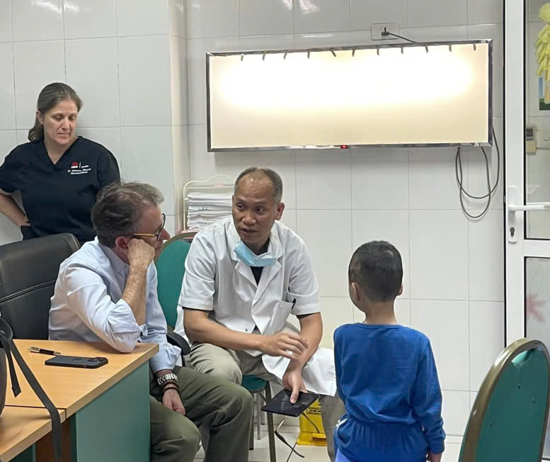
Patient 11
This 12 years old boy has his right ear reconstructed by using Medpor earframe. All were done in just one and only single-staged surgery using endoscopy assistant. 6 months later, the result is amazing. His friends don't even remember about his previous ear deformity anymore. He's very happy cuz' of this new buddy, he can wear glasses. This is a very realistic example of the recommendation that parent should have their children had new ear as soon as possible. Especially when in summer vacation or when you and your child have time for better furthermore developed social life.


Patient 12
The young boy, after undergoing one-stage ear reconstruction surgery using artificial cartilage, happily started the new school year — a big change marked by his new, beautifully shaped ear with clearly defined contours, perfectly matching the other ear. 📩 Message us or call the hotline for detailed, confidential consultation. 📞 0866.900.800 – 0974.700.600
News

Pre- and Post-Operative Procedure for Microtia Reconstruction with Dr Nguyen Hong Ha
Pre-Operative Procedure (Before Surgery) 1. Preparation Phase (4–6 Weeks Before Surgery) 2. Medical Evaluation (1–7 Days Before Surgery) 3. Patient Preparation Notes Surgical Day (Operation Day) Procedure Overview Immediate Surgical Goals Patient Notes Post-Operative Care & Recovery Timeline 1. Hospital Stay (5–7 Days) What Happens What You Should Do 2 Weeks After Surgery What Happens What You Should Do 3–6 Weeks After Surgery What Happens What You Should Do 7 Weeks – 2 Months What Happens What You Should Do 3–9 Months What Happens What You Should Do 12 Months What Happens What You Should Do Key Recovery Principles

Comprehensive Ear Reconstruction with Associate Professor Dr. Nguyen Hong Ha
Microtia—a congenital condition where the external ear is underdeveloped or absent—can deeply affect a person’s appearance, confidence, and quality of life. Under the expert care of Associate Professor Dr. Nguyen Hong Ha , patients now have access to advanced, world-class Medpor ear reconstruction that delivers natural, durable results with minimal scarring. Dr. Ha is Head of the Department of Maxillofacial Surgery, Plastic and Aesthetic Surgery at Viet Duc University Hospital and brings over 20 years of experience , with advanced training in France, the United States, and the United Kingdom . He is a recognized leader in reconstructive surgery in Vietnam and Southeast Asia. Advanced Single-Stage Medpor Reconstruction Dr. Ha has pioneered an innovative single-stage Medpor ear reconstruction technique , allowing complete ear reconstruction in just one operation. Using a biocompatible Medpor framework and endoscopic-assisted surgery , this approach offers: The procedure can be safely performed in children as young as 3–4 years old , helping prevent social and psychological challenges before school age. Who Can Benefit? Dr. Ha’s services are suitable for: More Than Reconstruction Beyond restoring an ear, Dr. Ha’s work restores confidence, identity, and quality of life —for children entering school without fear of stigma, and for adults finally closing a lifelong chapter. With proven expertise, compassionate care, and a commitment to global surgical standards, Associate Professor Dr. Nguyen Hong Ha offers a life-changing solution for patients and families affected by microtia.

Medpor Ear Reconstruction for Microtia by Dr. Nguyen Hong Ha
Microtia is a congenital condition in which the external ear is underdeveloped or completely absent. Beyond affecting facial symmetry, microtia can have a profound psychological impact on children and adults alike. With advances in reconstructive surgery, Medpor ear reconstruction has become a reliable and effective option for restoring a natural-looking ear with long-term stability. Under the expertise of Dr. Nguyen Hong Ha , Medpor ear reconstruction offers patients a carefully planned, modern solution that prioritizes both aesthetics and safety. What Is Medpor Ear Reconstruction? Medpor ear reconstruction uses a biocompatible porous polyethylene framework that is sculpted to replicate the natural anatomy of the ear. This framework is placed beneath the skin and gradually integrates with surrounding tissue as blood vessels grow into the material, creating a strong and stable reconstructed ear. Unlike traditional rib cartilage reconstruction, Medpor does not require harvesting cartilage from the patient’s chest , reducing donor-site pain and scarring. Key Advantages of Medpor Ear Reconstruction Medpor reconstruction offers several important benefits: These advantages make Medpor an appealing option for both pediatric and adult patients with microtia or anotia. Who Is a Suitable Candidate? Patients who may benefit from Medpor ear reconstruction include: A thorough consultation with Dr. Nguyen Hong Ha is essential to assess anatomy, skin condition, and overall health before proceeding. The Surgical Approach by Dr. Nguyen Hong Ha Dr. Nguyen Hong Ha applies meticulous surgical planning and refined techniques to ensure optimal outcomes. The Medpor framework is custom-shaped to match the patient’s facial proportions and positioned precisely to achieve symmetry with the opposite ear. Special attention is given to: Postoperative follow-up is an integral part of the process, allowing for careful monitoring and guidance throughout healing. Recovery and Results After surgery, patients typically experience: At follow-up milestones (such as 3–6 months), the reconstructed ear demonstrates natural contours, stable projection, and good skin mobility , closely resembling a natural ear. More Than Reconstruction: Restoring Confidence Medpor ear reconstruction is not only about creating an ear—it is about restoring confidence, identity, and quality of life . For children, early reconstruction can help prevent social anxiety and support healthy emotional development. For adults, it offers closure and renewed self-assurance. With experience in reconstructive surgery and a patient-centered approach, Dr. Nguyen Hong Ha is committed to delivering outcomes that are both technically sound and deeply meaningful. Conclusion Medpor ear reconstruction represents a modern, effective solution for patients with microtia or ear loss. When performed by an experienced reconstructive surgeon like Dr. Nguyen Hong Ha , the procedure offers reliable results, natural aesthetics, and lasting confidence—often in a single operation.

Congenital Ear Deformities in Children: Common Types, Causes, and Impact
Congenital deformities of the outer ear (auricle) are structural abnormalities present from birth that affect how the ear looks and functions. While not all ear deformities compromise health, they can have significant aesthetic and psychological effects on children , making early understanding and care essential for families. What Are Congenital Ear Deformities? During early pregnancy, the structures of the ear begin forming from the pharyngeal arches. By around week 20 of gestation , the external ear (auricle) normally reaches its full structural form. However, when development is disrupted, children may be born with deformities of the outer ear. Congenital ear deformities refer to a range of abnormal shapes or incomplete formation of the outer ear. These can affect one or both ears and vary in severity. Common Types of Ear Deformities Some typical congenital ear conditions include: These structural differences can influence not only appearance but also ear function. Causes of Ear Deformities The exact reasons for congenital ear deformities are often multifactorial, involving: Impact on Children Although visible ear deformities do not always affect overall health, they can have several consequences: 1. Aesthetic Concerns and Confidence Children with noticeable ear differences may feel self-conscious or socially isolated. These aesthetic differences can affect facial symmetry and draw unwanted attention, potentially impacting self-esteem. 2. Hearing Difficulties Some ear deformities interfere with the outer ear’s ability to capture sound effectively, potentially leading to reduced hearing on the affected side. Early hearing challenges can delay speech and language development . 3. Psychological and Social Effects Children with noticeable ear differences may encounter teasing or exclusion by peers, leading to anxiety or social withdrawal. Support from family and educators plays an important role in helping children adapt and thrive. 4. Increased Risk of Infection Abnormal ear anatomy may make the ear more susceptible to infections due to reduced natural protective structures. Diagnosis and Treatment Congenital ear deformities are usually identified at birth during routine newborn examinations. Diagnosis may involve physical assessment and, if needed, imaging or hearing tests. Treatment options depend on the type and severity of the deformity as well as the child’s age. Common approaches include: Early evaluation by a qualified specialist ensures tailored care for the best functional and cosmetic outcomes. Conclusion Congenital ear deformities range from mild cosmetic differences to more complex structural challenges. Understanding the types, causes, and effects of ear malformations helps parents and caregivers support children’s health, development, and confidence. With timely assessment and appropriate care, many children go on to lead healthy, fulfilling lives.